
Written By: Gary Hite, Research Content Writer
Reviewed By: Natalie Kunsman, M.D., Board-Certified Physician
Last Reviewed: September 10, 2026
Human chorionic gonadotropin (HCG) is one of the most extensively glycosylated glycoprotein hormones studied in the laboratory, and its glycosylation patterns sit at the center of how researchers interpret structure, heterogeneity, and analytical behavior. For laboratories working with HCG research samples, characterizing these carbohydrate structures is a foundational step in confirming identity, documenting batch consistency, and supporting reproducible experimental design. This article examines the glycosylation architecture of HCG and the analytical techniques used to map it, written for analytical chemists, biochemists, and academic researchers.
Disclaimer: HCG research samples are intended strictly for laboratory research and educational use only. They are not for human consumption and not for any diagnostic or therapeutic application. The information below describes analytical characterization for qualified researchers and academic professionals.
Why Glycosylation Defines HCG Sample Characterization
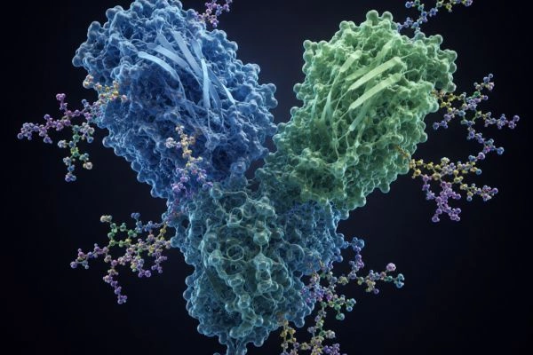
HCG is a heterodimeric glycoprotein built from two non-covalently associated subunits: a common alpha subunit and a hormone-specific beta subunit. Both subunits carry carbohydrate chains, and these glycans account for a substantial portion of the molecule’s total mass. Because the protein backbone is relatively conserved, much of the structural and analytical variability researchers observe in HCG samples traces directly back to glycosylation. For any laboratory characterizing HCG research material, mapping these carbohydrate structures is the most direct route to confirming molecular identity and documenting sample heterogeneity.
Glycosylation is also where HCG diverges into distinct molecular forms. Research samples can contain regular HCG, hyperglycosylated variants, free subunits, and partially degraded species, each carrying its own glycan signature. Reliable characterization depends on resolving these forms rather than treating the sample as a single uniform entity.
The Glycosylation Architecture of HCG
HCG carries both N-linked and O-linked glycans distributed across its two subunits. The molecule presents a defined set of glycosylation positions that analytical workflows aim to resolve site by site:
- Alpha subunit: two N-linked sites at asparagine residues Asn52 and Asn78.
- Beta subunit, N-linked: two sites at Asn13 and Asn30.
- Beta subunit, O-linked: four sites clustered on the C-terminal peptide region at serine residues Ser121, Ser127, Ser132, and Ser138.
N-linked glycans on HCG are predominantly complex biantennary structures, frequently capped with terminal sialic acid (N-acetylneuraminic acid) residues. O-linked glycans are typically mucin-type core 1 and core 2 structures, also commonly sialylated. The degree and pattern of sialylation is one of the most analytically significant features of an HCG sample, since it strongly influences apparent charge, chromatographic behavior, and characterized biochemical properties studied in research models, including in-vitro stability.
Hyperglycosylated HCG represents a distinct glycoform of research interest. It carries larger, more highly branched N-glycans (including triantennary structures) and more extended O-glycan chains than regular HCG. Distinguishing hyperglycosylated forms from regular HCG is a common analytical objective, and it depends almost entirely on detailed glycan profiling.
Sample Preparation and Glycan Release
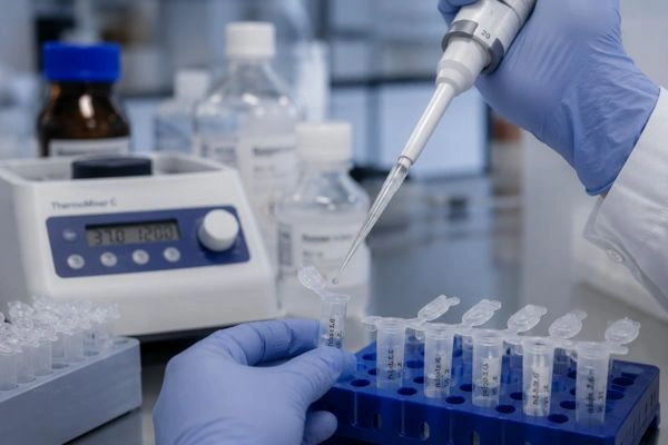
Effective glycan characterization begins with controlled release of the carbohydrate structures from the protein backbone. For N-linked glycans, researchers typically use the enzyme PNGase F, which cleaves the bond between the innermost N-acetylglucosamine and the asparagine residue, releasing the intact N-glycan pool. For O-linked glycans, which lack a single broadly specific releasing enzyme, chemical release through reductive beta-elimination is the standard approach. Selecting the correct release strategy at the outset determines which glycan population a given workflow will characterize.
Released glycans are often fluorescently labeled, commonly with 2-aminobenzamide (2-AB) or comparable tags, to enable sensitive detection during separation and to support relative quantification across samples. Labeling is a practical step that improves downstream resolution and reproducibility.
Mass Spectrometry for Glycan Profiling
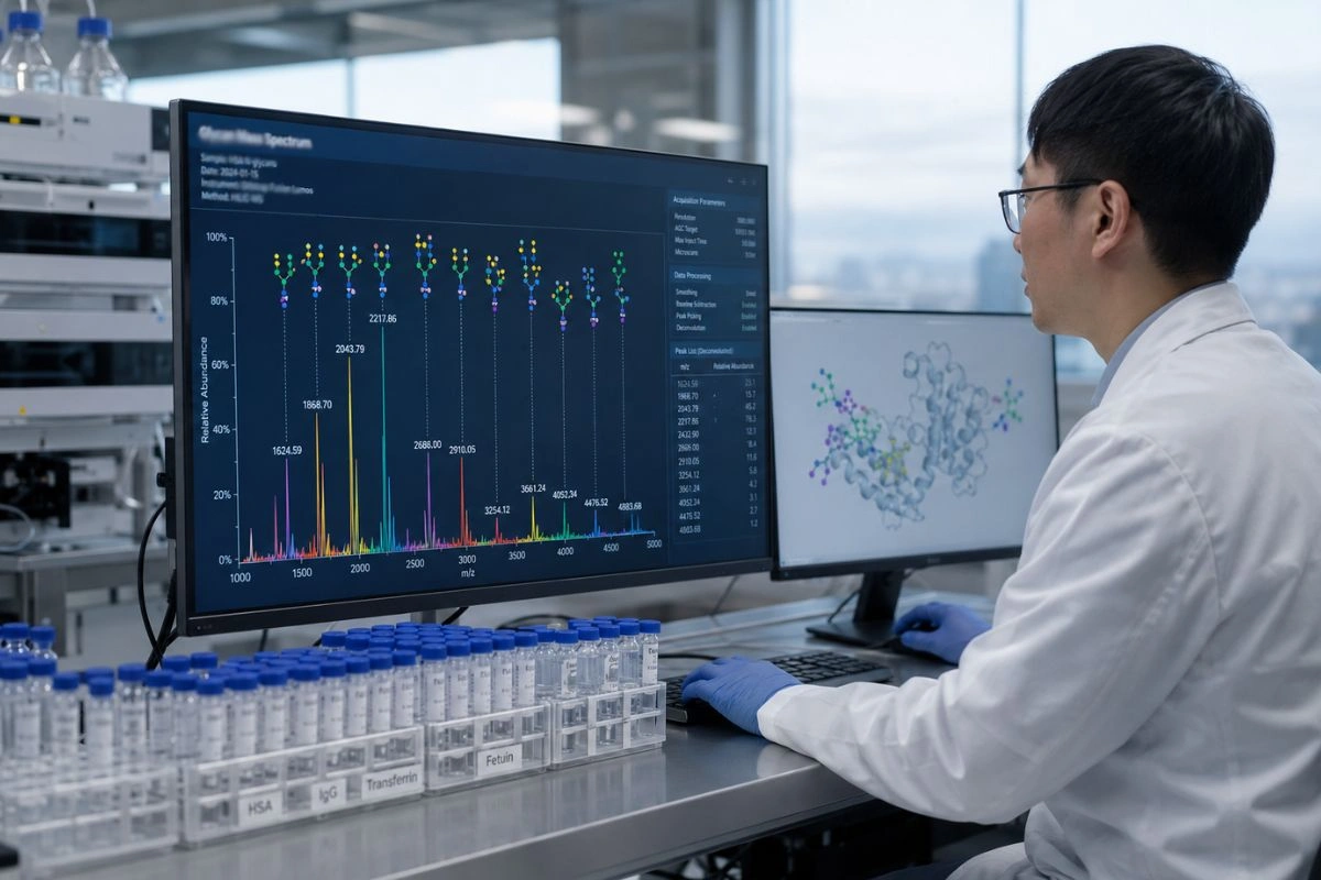
Mass spectrometry is the central technique for detailed HCG glycan characterization, and researchers apply it at several levels. Released glycan analysis profiles the pool of liberated N- or O-glycans, assigning compositions based on accurate mass. Matrix-assisted laser desorption/ionization time-of-flight (MALDI-TOF) MS and electrospray ionization (ESI) MS both serve this purpose, generating glycan mass profiles that reveal composition and relative abundance.
Glycopeptide analysis, often performed with liquid chromatography coupled to tandem mass spectrometry (LC-MS/MS), preserves the connection between a glycan and its specific attachment site. This bottom-up approach lets researchers assign glycan structures to individual sequons such as Asn52 or Asn30, producing site-specific glycosylation maps rather than a bulk average.
Intact and subunit-level mass spectrometry examines the assembled glycoprotein or its separated subunits, capturing the overall glycoform distribution and the macroheterogeneity of the sample. Combining these levels, released glycan, glycopeptide, and intact analysis, gives a layered and internally consistent picture of HCG glycosylation.
Chromatographic and Electrophoretic Separation
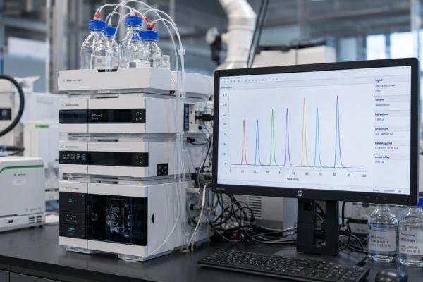
Separation science complements mass spectrometry by resolving glycan structures and glycoforms before or alongside detection. Hydrophilic interaction liquid chromatography (HILIC), frequently run at ultra-high-performance scale, separates labeled glycans according to size, composition, and linkage, and it pairs effectively with fluorescence detection and online MS. HILIC glycan mapping is a workhorse method for generating reproducible, comparable glycan profiles across HCG research batches.
Capillary electrophoresis with laser-induced fluorescence detection (CE-LIF) offers high-resolution separation of charged and labeled glycans, and it is particularly useful for resolving sialylated structures that differ in charge. Anion-exchange and other charge-based separations similarly help researchers profile sialylation patterns.
For monosaccharide composition, high-performance anion-exchange chromatography with pulsed amperometric detection (HPAEC-PAD) quantifies the individual sugar building blocks present in a sample, while dedicated sialic acid assays quantify terminal N-acetylneuraminic acid content. These compositional measurements anchor the more detailed structural data.
Linkage Analysis, Lectins, and NMR
Beyond composition and profiling, several techniques address fine structural detail. Exoglycosidase sequencing uses panels of specific enzymes to remove monosaccharides one linkage at a time, and the resulting shifts in chromatographic or electrophoretic profiles confirm both the identity and the linkage of terminal residues. Researchers apply this approach to verify structures proposed from mass data.
Lectin-based methods, including lectin affinity chromatography and lectin microarrays, exploit the selective binding of lectins to particular glycan motifs. These tools support rapid screening and comparative profiling of glycan features across HCG samples.
Nuclear magnetic resonance (NMR) spectroscopy provides the most detailed structural information, resolving anomeric configurations, linkage positions, and monosaccharide identities. While NMR requires larger sample amounts and specialized expertise, it serves as a definitive reference method for fully characterizing HCG glycan structures.
Managing Glycoform Heterogeneity in Research Samples
Glycosylation makes HCG inherently heterogeneous, and characterizing that heterogeneity is often the central analytical goal rather than an obstacle to it. Microheterogeneity refers to the range of glycan structures occupying a single site, while macroheterogeneity refers to variation in site occupancy across the molecule. A thorough characterization documents both, since each contributes to the observed glycoform distribution. Researchers establishing reference characterization for an HCG sample typically report site-specific glycan structures, relative abundances, sialylation patterns, and the proportion of any hyperglycosylated or free-subunit species present.
This level of documentation supports reproducibility. When glycan profiles are recorded consistently, laboratories can compare batches, monitor stability over storage, and ensure that experimental results rest on well-defined material. International reference preparations for HCG have themselves been characterized through extensive glycan analysis, illustrating how central glycosylation profiling is to defining the molecule.

Building a Reliable Characterization Workflow
To characterize an HCG research sample effectively, combine orthogonal techniques rather than relying on a single method. A practical workflow starts with compositional and intact-level measurements to establish the broad glycoform picture, proceeds to released glycan profiling by HILIC and mass spectrometry, and adds glycopeptide LC-MS/MS for site-specific assignment. Exoglycosidase sequencing or NMR then confirms fine structural detail where definitive assignment is required. Cross-checking results across these independent approaches strengthens confidence in the final characterization and reduces the risk of misassignment.
Documenting methods, instrument parameters, and data processing settings is equally important. Reproducible glycan characterization depends not only on the techniques chosen but on consistent, well-recorded protocols that other researchers can follow and verify.
Conclusion
Glycosylation patterns define much of what makes HCG analytically distinctive, from its subunit-specific N- and O-glycan sites to the sialylation features and hyperglycosylated variants that separate one glycoform from another. Characterizing these structures relies on a coordinated toolkit: controlled glycan release, mass spectrometry at released, glycopeptide, and intact levels, high-resolution chromatographic and electrophoretic separation, and confirmatory linkage analysis by exoglycosidase sequencing or NMR. For analytical chemists and academic researchers, mapping HCG glycosylation with orthogonal methods delivers the structural clarity and reproducibility that rigorous research requires.
Disclaimer: All products and reference materials referenced are intended strictly for laboratory research and educational use only. HCG research samples are not for human consumption, are not for diagnostic or therapeutic use, and are not intended to treat, cure, or prevent any condition. Nothing in this article constitutes medical, clinical, or professional advice. This content is provided for informational and educational purposes for qualified researchers and academic professionals. Handling, storage, and use of research materials must comply with all applicable institutional guidelines, safety protocols, and local, state, and federal regulations.
Frequently Asked Questions
Why does glycosylation matter when characterizing HCG research samples?
Because the HCG protein backbone is relatively conserved, most of the structural variability you observe traces back to its carbohydrate chains. Mapping these glycans is the most direct way to confirm molecular identity, document batch consistency, and resolve the distinct molecular forms a sample may contain, including regular HCG, hyperglycosylated variants, and free subunits. Approach glycan profiling as a foundational step rather than an optional add-on.
How many glycosylation sites does HCG carry?
HCG presents a defined set of sites across both subunits. The alpha subunit holds two N-linked sites at Asn52 and Asn78. The beta subunit adds two N-linked sites at Asn13 and Asn30, plus four O-linked sites clustered on the C-terminal peptide region at Ser121, Ser127, Ser132, and Ser138. Target each position individually when building a site-specific glycosylation map.
What distinguishes hyperglycosylated HCG from regular HCG?
Hyperglycosylated HCG carries larger, more highly branched N-glycans, including triantennary structures, along with more extended O-glycan chains than regular HCG. Distinguishing the two glycoforms depends almost entirely on detailed glycan profiling, so apply released glycan analysis and glycopeptide mapping rather than relying on intact mass measurements alone.
Which analytical techniques work best for mapping HCG glycans?
Combine orthogonal methods for the most reliable results. Use mass spectrometry at released, glycopeptide, and intact levels to capture composition and site occupancy, pair HILIC and CE-LIF for high-resolution separation of labeled and sialylated structures, and confirm fine structural detail with exoglycosidase sequencing or NMR. No single technique delivers a complete picture on its own, so cross-check assignments across independent approaches.
How are glycans released from HCG before analysis?
Select your release strategy based on the glycan population you want to characterize. For N-linked glycans, use the enzyme PNGase F to cleave the intact N-glycan pool from the protein backbone. For O-linked glycans, which lack a single broadly specific releasing enzyme, apply chemical release through reductive beta-elimination. Fluorescently label the released glycans, commonly with 2-AB, to improve detection sensitivity and reproducibility downstream.